
DENTAL X – RAY : –
Dental x-ray shows :
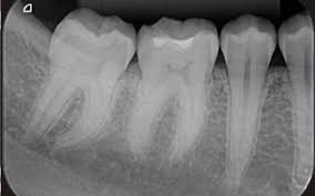
Cavities, spescially small areas of decay between teeth.
Decay beneath existing filling.
Bone loss in your jaw.
Areas of infection.
The position of unerupted or impacted teeth.
Abscessed teeth ( infection at the root of your tooth or between your gums and your tooth )
Cysts and some type of tumors.
WHAT IS THE PRINCIPLE OF X-RAY IN DENTISTRY : –
The technique is based on the principle of aiming the central ray of the x-ray beam at 90° to an imaginary line which bisects the angle formed by tha long axis of the tooth and the plane of the receptor. The image receptor is placed as close as possible to the tooth under investigation, without bending the packet.
WHAT ARE THE MAIN PARTS OF THE DENTAL X-RAY UNIT : –
1 . The tube head,
2 . An extension arm,
3 . The control panel.
WHAT ARE THE USES OF X-RAY IN DENTISTRY : –
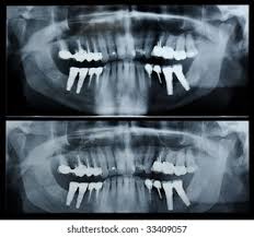
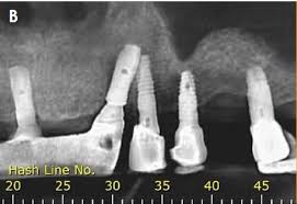
It is used to plan treatment for dental implants, check for impacted wisdom teeth and detect jaw problems.
A panoramic x-ray in not the best method for detecting cavities, unless the decay is very advanceed and deep.
WHAT ARE 3 COMMON USES OF X-RAYS : –
It can help to find broken bone, tumors and foreign objects in the body.
It also used in other types of examination and procedure, including CT-SCANS, MAMMOGRAMS, FLUOROSCOPY.
WHAT ARE THE TYPES OF X-RAYS : –
Plain x-ray,
Computed tomography,
Fluoroscopy – which produces moving images of organs,
Mammography :- An x-ray of the breasts.
Angiography :- An x-ray of blood vessels.
HOW MANY DENTAL X-RAYS ARE SAFE : –
Most people get an average of 20 x-rays per year and are always safe.
WHAT TYPE OF X-RAY IS DENTAL : –
It is 2 type.
1 . Intraoral,
Extraoral.
Each type serves specifice diagnostic purpose and provides different views of the mouth’s internal structures.
HOW ARE X-RAY PRODUCED IN THE DENTAL CLINIC : –
X-rays are created inside the x-ray head. Electrical current passes between the anode and the cathode and hit the target areas where x-rays are produced. The x-rays then travel through the positioning indicator device where the x-ray beam exposes the receptor.
WHAT ARE DENTAL X-RAYS MEASURED IN : –
It is the GRAY ( Gy ), but in dental radiograph the dose lavels are usually a small fraction of 1 Gy milliGray ( mGy ) or even micro Gray.
WHAT EQUIPMENT IS USED FOR X-RAYS :-
An x-ray equipmant is consists of 4 main parts,
The x-ray tube,
The transformer,
The control panal,
The tube stand.
The tube emits x-rays, which get absorbed by your soft tissues, bones and other structures when they enter the body, thus appearing white on the film.
WHAT IS THE BASIC PRINCIPLE OF X-RAY : –
The reduction of energy is caused by absorption, which is the main principle of x-ray imaging. X-ray radiography measures the amount of energy loss. Because this energy loss differs for the different material, we can see a certain contrast in the image.
WHAT ARE THE 4 MAIN COMPONENTS OF THE X-RAY :-
A Cathode,
An Anode,
A vaccum enclosure,
4.High voltage cable forming the x-ray generator attached to the cathod and anode componants.
DENTAL X-RAY STEPS :-
Greet your patient and guide them to their exam room.
Ensure the patient is comfortable before you proceed.
Place the lead apron and thyroid collar around your patient.
Incert the image sensor into the patient’s mouth.
Instruct the patient to hold the position and not move.
Then shoot the x-ray from the control panel.
PROPERTIES OF X-RAYS :-
They have a shorter wavelength of the electromagnatic spectrum.
Requires high voltage to produce x-rays.
They are used to capture the human skeleton diffects.
They travel in a straight line and donot carry an electric charge with them.
They are capeble of travelling in a vaccum.
WHAT IS THE FUL FORM OF X-RAY :-
X-ray is a form of electromagnetic radiation. X-rays were discovered in 1895 by Wilhelm Conrad Rontgen, who named the few form of radiation X-radiation ( ‘X’ standing for unknown ). X-radiation is called Rontgen radiation, after Wilhelm Rontgen.
HOW TO IDENTIFY FRACTURES IN X-RAY :-
It diagnose bone fractures by showing a darkar line or gap where the bone is broken.
WHAT ARE THE BENEFITS OF X-RAYS :-
Diagnose possibly life threatening condition such as blocked blood vessels, bone cancer and infections.
TOP 5 USES OF X-RAYS :-
To evaluate the symptoms of the body.
To diagnose injuries.
To perform dental check-ups.
To diagnose cancer.
To identify joint changes.
WHAT IS A DENTAL X-RAY CALLED :-
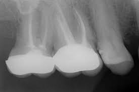
Periapical x-rays.
A periapical x-ray shows entire tooth starting from the crown to the root tip. It help your dentist to diagnose tooth decay, gum disease, bone loss or any other abnormalities in your tooth and surrounding Bone and tissues.
HOW LONG DO X-RAYS STAYED IN THE BODY :-
After a radiography or ct scane no radiation remains in the body.
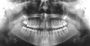
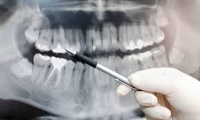
WHAT IS OPG IN DENTAL :-
OPG or Orthopentomogram is special x-ray of the lower face, teeth and jaw. An OPG is a panaromic view of x-ray of the lower face and jaw which displays allthe teeth of upper and lower jaw on a single film.
WHAT IS THE FULL FORM OF RVG :-
RVG stands for RADIO VISIO GRAPHY digital x-ray.
WHAT IS THE COST OF OPG :-
They canbe anywhere 350/- to 550/-.
WHICH ARE THE 3 PILLARS OF X-RAY PROTECTION :-
1 . Time
2 . Distance
3 . Shielding
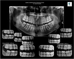
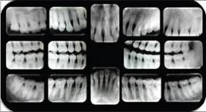
HOWMANY X-RAY IN A FULL MOUTH SET :-
An FMX seriese of 18-20 x-rays which includes all 4 bitewings and PA’s of every tooth.